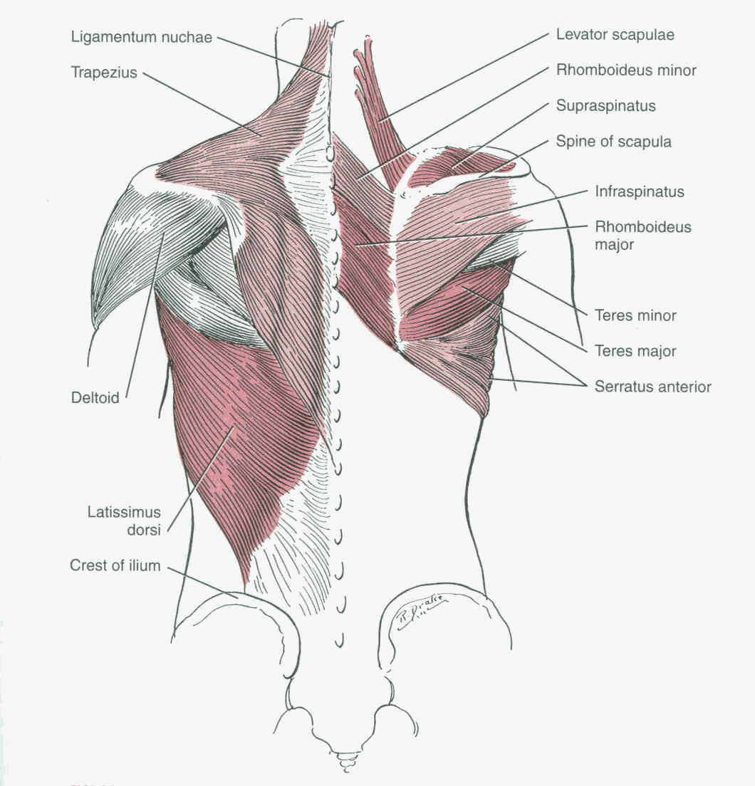
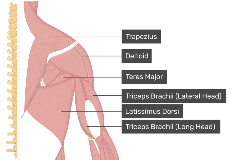
Shoulder Muscles Diagram : The muscles of the shoulder support and produce the movements of the shoulder girdle.they attach the appendicular skeleton of the upper limb to the axial skeleton of the trunk.. Plus, exercises for training them. The muscles of the shoulder support and produce the movements of the shoulder girdle.they attach the appendicular skeleton of the upper limb to the axial skeleton of the trunk. The shoulder muscles are a set of complex muscles that act as a link between the torso and the head or neck. These muscles form the outer shape of the shoulder and underarm. The shoulder muscles and shoulder tendons involved with shoulder mobility include the four rotator cuff muscle and tendon pairs:
Shoulder muscle anatomy neck muscle anatomy shoulder muscles supraspinatus muscle muscle fascia muscle diagram human body organs anatomy images latissimus dorsi. Subscapularis, supraspinatus, infraspinatus and teres minor. The rotator cuff is a group of four muscles and tendons that surround the glenohumeral joint. The muscles of the shoulder support and produce the movements of the shoulder girdle.they attach the appendicular skeleton of the upper limb to the axial skeleton of the trunk. The shoulder muscles consist of the deltoids and the rotator cuff group.the deltoids are the muscles that can be seen on the outside of the body, whilst the rotator cuff group is found within the shoulder joint itself, providing structural support and allowing the shoulder to perform many functions.

Related posts of shoulder muscles and tendons diagram muscle anatomy back.
Bones in shoulder, ligaments of the shoulder joint, parts of the shoulder joint, shoulder anatomy, shoulder joints and muscles, shoulder structure anatomy, shoulder tendon anatomy, shoulder tendons ligaments, human muscles, bones in shoulder, ligaments of the shoulder joint, parts of. Related posts of shoulder muscles and tendons diagram muscle anatomy back. Muscles of the shoulder : The most common shoulder injuries are sprains, strains, and tears. The most common shoulder injuries are sprains, strains, and tears. Learn about these muscles, their origin and insertion points, and their functional anatomy. Four of them are found on the anterior aspect of the shoulder, whereas the rest are located on the shoulder's posterior aspect and in the back. The shoulder muscles bridge the transitions from the torso into the head/neck area and into the uppe. The bursa is a small sac of fluid that cushions and. The large deltoid muscle is the outer layer of shoulder muscle. What are common rotator cuff injuries? Subscapularis, supraspinatus, infraspinatus and teres minor. This flexibility is also what makes the shoulder prone to instability and injury.
Learn about these muscles, their origin and insertion points, and their functional anatomy. Bones in shoulder, ligaments of the shoulder joint, parts of the shoulder joint, shoulder anatomy, shoulder joints and muscles, shoulder structure anatomy, shoulder tendon anatomy, shoulder tendons ligaments, human muscles, bones in shoulder, ligaments of the shoulder joint, parts of. For that reason, and because of the dexterity of the shoulder joint itself, the musculature of the shoulder is complex, ranging from massive prime mover muscles to finer stabilizer and fixator muscles. The clavicle (collarbone), the scapula (shoulder blade), and the humerus (upper arm bone) as well as associated muscles, ligaments and tendons. These muscles form the outer shape of the shoulder and underarm.

The shoulder blade (scapula) connects to the collarbone (clavicle) at this joint.
Related posts of shoulder muscles and tendons diagram muscle anatomy back. The shoulder muscles are associated with movements of the upper limb shoulder muscles diagram. The most common shoulder injuries are sprains, strains, and tears. The main shoulder muscles are trapezius, deltoid, pectoralis major and 4 rotator cuff muscles: The rotator cuff is a group of four muscles and tendons that surround the glenohumeral joint. And the ligaments, which connect bones. The partner should slowly, but firmly press on both sides of your shoulder to compress the ac joint. Muscle anatomy back 12 photos of the muscle anatomy back back muscle anatomy images, back muscle anatomy of the human body, back pain muscle anatomy, muscle anatomy lower back, posterior back muscle anatomy, human muscles, back muscle anatomy images, back muscle anatomy of the human body, back pain muscle anatomy. Shoulder muscle anatomy neck muscle anatomy shoulder muscles supraspinatus muscle muscle fascia muscle diagram human body organs anatomy images latissimus dorsi. Diagramme schnell und einfach erstellen. The rotator cuff muscles are important stabilizers and movers of the shoulder joint. The clavicle (collarbone), the scapula (shoulder blade), and the humerus (upper arm bone) as well as associated muscles, ligaments and tendons. Numerous muscles help stabilize the three joints of.
Shoulder joint of human body anatomy infographic diagram with all parts including bones ligaments muscles bursa cavity capsule cartilage membrane for medical science education and health care. Shoulder muscle anatomy neck muscle anatomy shoulder muscles supraspinatus muscle muscle fascia muscle diagram human body organs anatomy images latissimus dorsi. And the ligaments, which connect bones. The following is an overview of the shoulder muscle anatomy. The tendons, which anchor muscle to bone;

The muscles of the shoulder support and produce the movements of the shoulder girdle.they attach the appendicular skeleton of the upper limb to the axial skeleton of the trunk.
Cuff muscles of the back shoulder muscles and chest human anatomy diagram the shoulder muscles bridge the transitions. Diagram of the shoulder, including the location of the rotator cuff. As one of the four muscles of the rotator cuff, the main function is to externally rotate the humerus and stabilize the shoulder joint. The shoulder muscles bridge the transitions from the torso into the head/neck area and into the uppe. This often happens when stress is placed on the tissues that stabilize the shoulder—the muscles; The humeral head in the glenoid socket. The deltoid is the largest, strongest muscle of the shoulder. The rotator cuff muscles are important stabilizers and movers of the shoulder joint. Muscle anatomy back 12 photos of the muscle anatomy back back muscle anatomy images, back muscle anatomy of the human body, back pain muscle anatomy, muscle anatomy lower back, posterior back muscle anatomy, human muscles, back muscle anatomy images, back muscle anatomy of the human body, back pain muscle anatomy. The articulations between the bones of the shoulder make up the shoulder joints.the shoulder joint, also known as the glenohumeral joint, is the major joint of the shoulder, but can more broadly include the. The large deltoid muscle is the outer layer of shoulder muscle. The shoulder muscles are associated with movements of the upper limb shoulder muscles diagram. Plus, exercises for training them.

0 Komentar